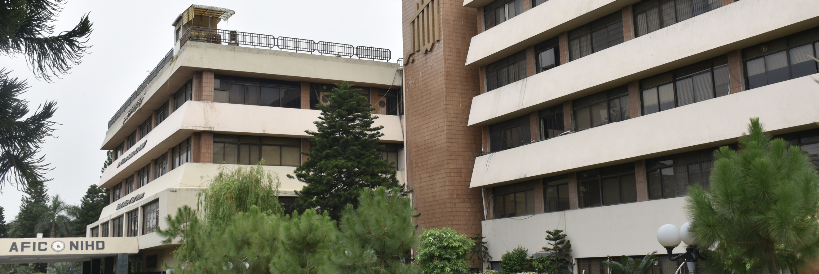
February 2013
TRUNCUS ARTERIOSIS TYPE-II (First successful surgery of Pakistan)
Truncus Arteriosis is a normal feature in an amphibians like frogs while it is a congenital anomaly in human beings. It usually presents as emergence of a single common arterial trunk from left ventricle. Persistent Truncus Arteriosis is 1% of all congenital heart defects.
In Pakistan, the first successful surgery was done at AFIC/NIHD on 29 Nov 12 when 8 months old patient Eshal resident of Swat was operated by a dedicated team of AFIC/NIHD doctors. The patient was suffering from Truncus Arteriosis Type-II in which her both Pulmonary arteries were originating independently from truncal aorta and having sub aortic VSD (hole) in ventricle. During planned surgical procedure, patient went through artificial cardio pulmonary by-pass , the hole was closed with synthetic patch, the pulmonary arteries were removed from truncal aorta , the aorta closed directly , animal conduit made from bovine pericardium known as contegra conduit was interposed between right ventricle and pulmonary confluence . The patient responded very effectively to post operative care/ treatment and discharge on 24 Dec 2012 with an excellent prognosis.
Col Kamal Saleem
FCPS (Surgery), FCPS (Cardiac Surgery)
Consultant Paediatric Cardiac surgeon
December 2012
PTMC – in a small child with pulmonary balloon
A 05-year-old male child, weighing 11 kg, presented with progressive exertional dyspnea (NYHA III) for the last few months. Clinical examination revealed signs of CCF, loud S1 & P2, opening snap and apical mid diastolic rumbling murmur.
Echocardiography revealed thickened mitral leaflets, MVA = 0.4 cm2, MPG = 22 mmHg, non–calcified MV and there was no clot in LA. He was diagnosed as a case of severe mitral stenosis of rheumatic etiology and managed with anti-failure medication for one week.
PTMC was planned after stabilization. The maximum balloon for PTMC we could use for this size child was 16mm and which was not available in inoue; so we decided to use the pulmonary balloon (VACS II- 16 mm). After septal puncture with standard technique, we advanced Mullin sheath into LA over loopy wire, then Mitral valve crossed with balloon wedge catheter (6Fr) through the same sheath and exchanged with super stiff exchange wire (0.35’’) Successful PTMC was done with VACS II Pulmonary Balloon under echocardiography guidance.
Post procedural angiographic gradient between LA to LV dropped from 18mmhg to 6mm hg and echocardiography revealed MPG of 08 mmHg, MVA 1.60 cm2 with MR of less than grade I. He was discharged next day and follow up after 2 weeks showed remarkable improvement in his symptoms. Now the patient is on regular follow up and doing well.
To the best of our knowledge, this is the first reported PTMC in a youngest child with rheumatic Mitral Stenosis weighing only 11 kgs.
Brig. Dr. Maad Ullah
MBBS, DHC, FCPS (Paeds)
MRCP (Peads), OJT (Paeds) Card UK
Paediaric Cardiologist
AFIC/NIHD Rawalpindi